Nervous System – Part I
The Nervous system is the most complex system in our body. Metaphorically, one can think of management system. The basic unit of it is cell, termed as neuron. (Fig. 1)
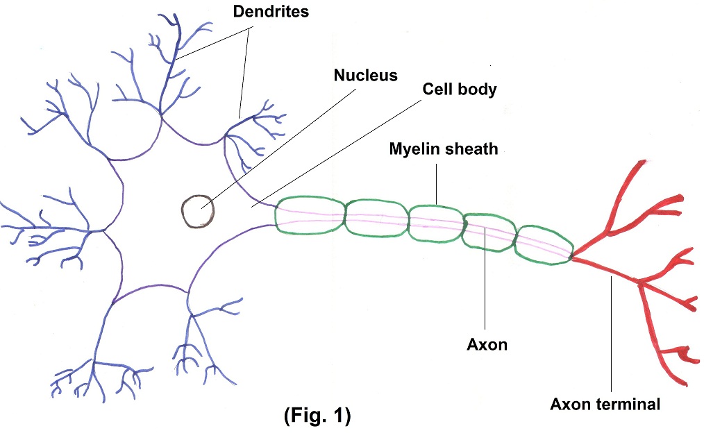
It consists of —
- Cell body – It contains cell organelle and nucleus. It is responsible for controlling major metabolic activities in neuron.
- Dendrites – This tree shaped structure is receiving portion of neuron. They contain numerous receptors for binding chemical signals from the cell.
- Axons – It is long and thin structure, which carry forward nerve impulse to another neuron or muscle fiber.
These specialized cells perform most of the unique functions like sensing, thinking, remembering, controlling muscle activity etc. As neurons differ in their length and shape, so they differ in their properties also. Neuroglias are smaller cells than neurons. It supports, nourishes and protects neurons.
How do these cells communicate between themselves and with external and internal environment? It is of two types.
- Electrical stimulation – Like muscle cells, neurons possess electrical excitability, that means, ability to respond to a stimulus and convert it into an action potential. Action potential begins and travels due to movement of ions (such as Na and potassium) e.g. direct cell to cell as occurs in cardiac muscles, smooth muscles of G. I. tract.
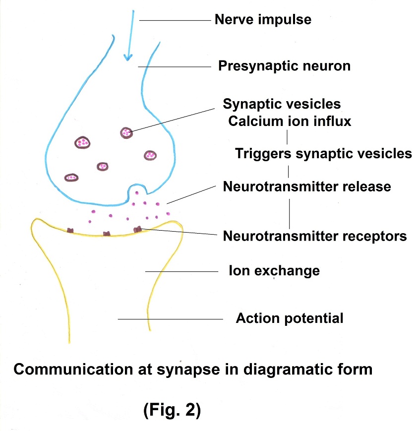
- Chemical stimulation – It occurs between two neurons or between neuron and an effector cell. The site of communication is synapse. The synaptic vesicle stores a chemical that is called as neurotransmitter. It is a molecule which excites or inhibits another neuron, muscle fiber or gland.
Together it is called as electro – chemical communication.
About 1000 Neurotransmitters are known. Out of that some are excitatory, and some are inhibitory. e.g. GABA — inhibitory neurotransmitters.
Communication at synapse in diagrammatic form. (Fig.2)
Let us study the organizational structure of this system. (Fig. 3)
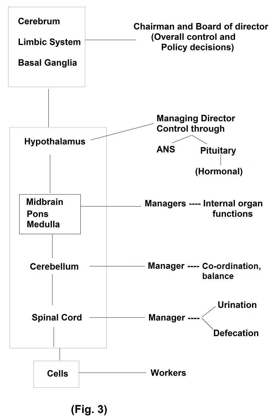
Cerebral cortex, limbic system and basal ganglia can be regarded as directors, receiving essential information through sensory pathways, taking major decisions and sending orders through motor pathways.
Executive work is vested to thalamus, hypothalamus and midbrain structures. Processing of information, maintaining internal balance while interacting with external environment occurs at this level.
The cells in other bodily organs and systems can be regarded as workers carrying out jobs given to them.
Now we will study components of Nervous system and then its functional organization at various levels.
The nervous system consists of two main parts, the Central Nervous System (CNS) and the Peripheral Nervous System (PNS).
Components of Nervous System –
The Central Nervous System contains the Brain and Spinal Cord.
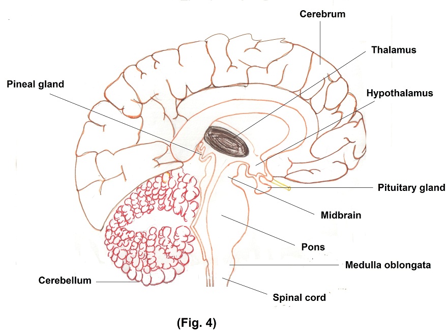
The main areas of the brain are – Fig. 4
- Cerebrum
- Diencephalon – Thalamus, Hypothalamus, Pineal gland
- Brainstem – Midbrain, Pons, Medulla Oblongata
- Cerebellum
Function —
The brain is the control centre for registering sensations, processing information, making decisions and sending orders to working organs. It is also a centre for intellect, emotions, behavior and memory.
Through these functions, nervous system maintains homeostasis (means physiological balance).
1. Cerebrum –
Cerebrum has four lobes — Frontal, Parietal, Temporal and Occipital. Basal ganglia help to regulate initiation and termination of movement. Limbic system is called as emotional brain because it plays a primary role in range of emotions for example – anger, pleasure, love etc.
i. Sensory areas in the cerebrum receive sensory information and are involved in conscious awareness of sensation that is called as perception.
ii. Motor areas control execution of voluntary movements.
iii. Association areas deal with more complex integrative functions like memory, emotions, reasoning etc.
Cerebrum is supported by diencephalon and brainstem.
2. Diencephalon –
It consist of –
i. Thalamus is a major relay station for most sensory inputs.
ii. Hypothalamus is a one of the major regulators of homeostasis with the help of pituitary gland which in turns controls hormonal balance. Hypothalamus monitors osmotic pressure, blood glucose level, hormone concentration etc. It also regulates body temperature. It controls and integrates functions of autonomic nervous system i.e. contraction of smooth muscles, cardiac muscles and secretions of many glands.
iii. Pineal gland or epithalamus – It secretes melatonin which promotes sleepiness.
3. Brainstem –
Brainstem is in continuation of spinal cord upwards. It consists of
i. Medulla – Various nuclei (control centers) of cardio–vascular system and respiratory centre is present in medulla. Therefore, injury to medulla may lead to death. All sensory and motor tracts pass through medulla. Cortico-spinal tract decussate (cross-over) at the level of medulla.
ii. Pons — It is a connecting bridge between medulla and midbrain. Pneumotaxic area is present in pons which helps to maintain breathing along with medulla.
iii. Midbrain — Several nuclei are present in midbrain. Substantia nigra present in midbrain releases dopamine, which controls subconscious motor activity. In Parkinson’s disease due to degenerative changes in this areas dopamine secretion decreases.
Reticular formation – It is a net like arrangement which extends from superior part of spinal cord through the brainstem into diencephalons i.e. thalamus and hypothalamus. Reticular activating system does not allow unnecessary information to go up and is responsible for consciousness, alertness and arousal from sleep.
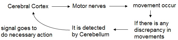
4. Cerebellum –
Primary function of cerebellum is to evaluate movements. e.g.
Spinal cord –
The spinal cord is pathway for sensory input to brain and motor output from the brain via spinal nerves. For certain sensory input the response occurs at the level of spinal cord e.g. spinal cord reflex. When we touch any hot object, we remove our hand immediately. This rapid action occurs because of this reflex. Autonomic control over urination and defecation occur at the level of spinal cord.
We will discuss Peripheral Nervous System (PNS) in second part.