Hearing and Ear
Hearing is the ability to perceive sounds. The ear is designed to collect and amplify sound and efficiently transfer the mechanical energy of sound waves into nerve impulse. The ear also contains receptors for equilibrium. (Fig. 1)
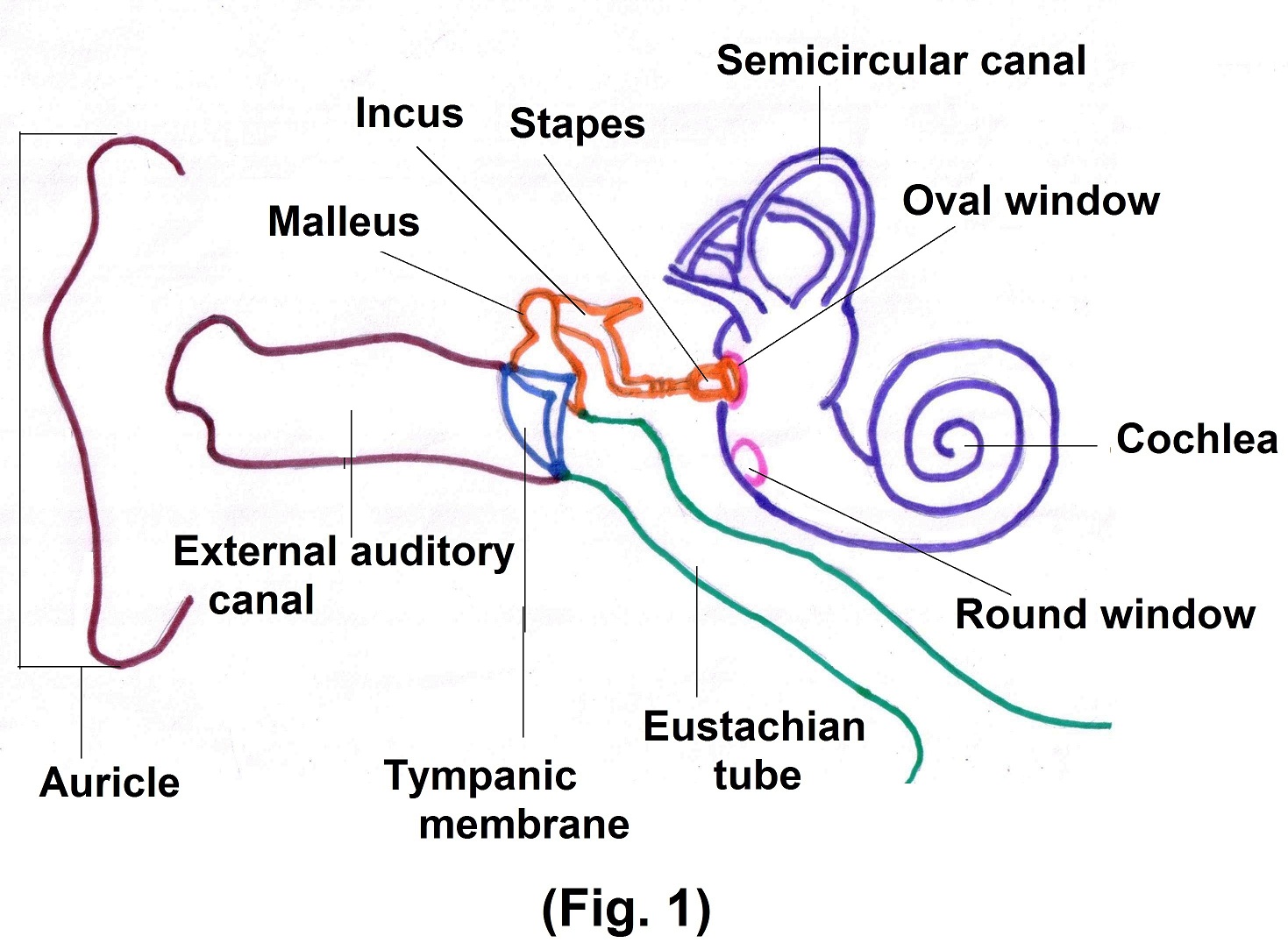
Ear is divided into 3 parts external ear, middle ear and internal ear. External ear consists of auricle, external auditory canal and eardrum. Middle ear consists of oval window, round window and three smallest bones incus, malleus and stapes called auditory ossicles.
The internal ear is also called the labyrinth. It consists of two main divisions, an outer bony labyrinth and inside membranous labyrinth. The bony labyrinth is a series of cavities in petrous portion of temporal bone, divided into three areas –
- semicircular canal
- vestibule
- cochlea
Bony Labyrinth contains perilymph, a fluid, which surrounds and protects membranous labyrinth. Membranous labyrinth is like long balloons put inside a rigid tube. Membranous labyrinth contains endolymph, a fluid which has a role in the generation of auditory signals. Auricle collects sound waves, while external auditory canal directs sound wave to ear drum. When sound waves strike tympanic membrane, alternating waves of high and low pressure in the air, cause the tympanic membrane to vibrate back and forth. It vibrates slowly with Low frequency sound wave and vibrates rapidly in response to high frequency sound.
The central area of tympanic membrane is connected to the malleus, which vibrates along with the tympanic membrane. This vibration is transmitted from malleus to incus and then to the stapes. As stapes moves back and forth its oval shaped foot plate which is attached via ligament to the circumference of oval window (a small opening in the bony partition that separate middle ear from internal ear) starts vibrating. The vibrations of oval window are about 20 times more vigorous than tympanic membrane.
The movement of stapes at the oval window sets up fluid pressure waves in the perilymph of the cochlea. Subsequently, pressure waves are transmitted to the vestibular membrane and then into the endolymph inside the cochlear duct. (Fig. 2)
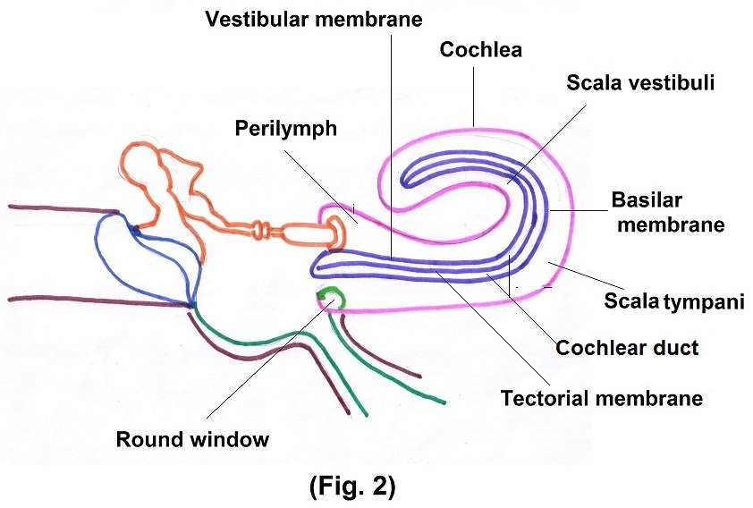
Pressure wave in endolymph, cause the basilar membrane (membrane that separates cochlear duct from the Scala tympani) to vibrate, which ultimately leads to the generation of nerve impulses in cochlear nerve fibers.
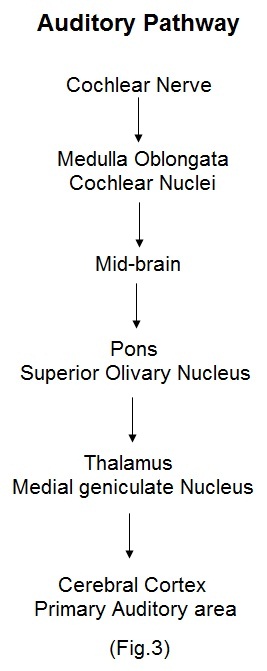
Axons of cochlear nerve synapse with neurons in the cochlear nuclei in the medulla oblongata on the same side. Some of the axons from cochlear nuclei, cross over in medulla, ascend in a tract, called the lateral meniscus, on the opposite side and terminate in midbrain. Other axons from the cochlear nuclei end in the superior olivary nucleus in the pons on each side. From pons, impulses are conveyed to the medial geniculate nucleus in the thalamus and finally to the primary auditory area of the cerebral cortex, where perception of hearing occurs. Because many auditory axons cross over in the medulla while others remain on same side, the right and left primary auditory area receive nerve impulses from both ears.
Suggested further reading for Section II –
PHYSIOLOGY —
- Guyton and Hall, Textbook of Medical Physiology, Philadelphia, PA: Saunders Elsevier.