Nervous System- Part II
Peripheral Nervous System (PNS) –
PNS consists of all nervous tissues outside CNS. It connects CNS (i.e. brain and spinal cord) to external and internal environment.
Components of PNS – (Fig.5)
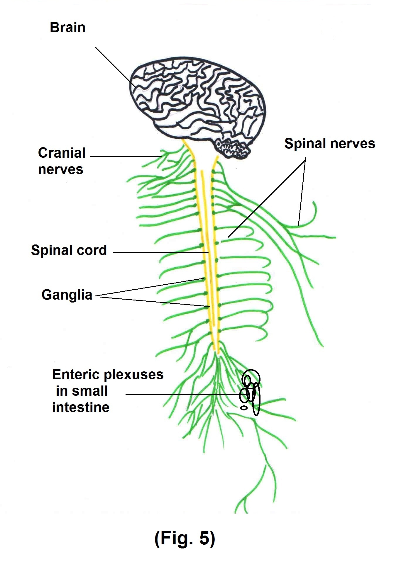
- Nerves – There are 12 pairs of cranial nerves that emerge from brain and their sensory neuron cause perception through special senses like hearing, vision etc. There are 31 pairs of spinal nerves that emerge from spinal cord and connect CNS to sensory receptors on skeletal muscles.
- Ganglia – It primarily consist of neuronal cell body that are located outside brain and spinal cord.
- Enteric plexus – It is an extensive network of nerves located in walls of organ of GIT.
- Sensory receptors – which monitor changes in external or internal environment, e.g. touch receptors in skin, photo receptors in eye etc.
As you see in the diagram –
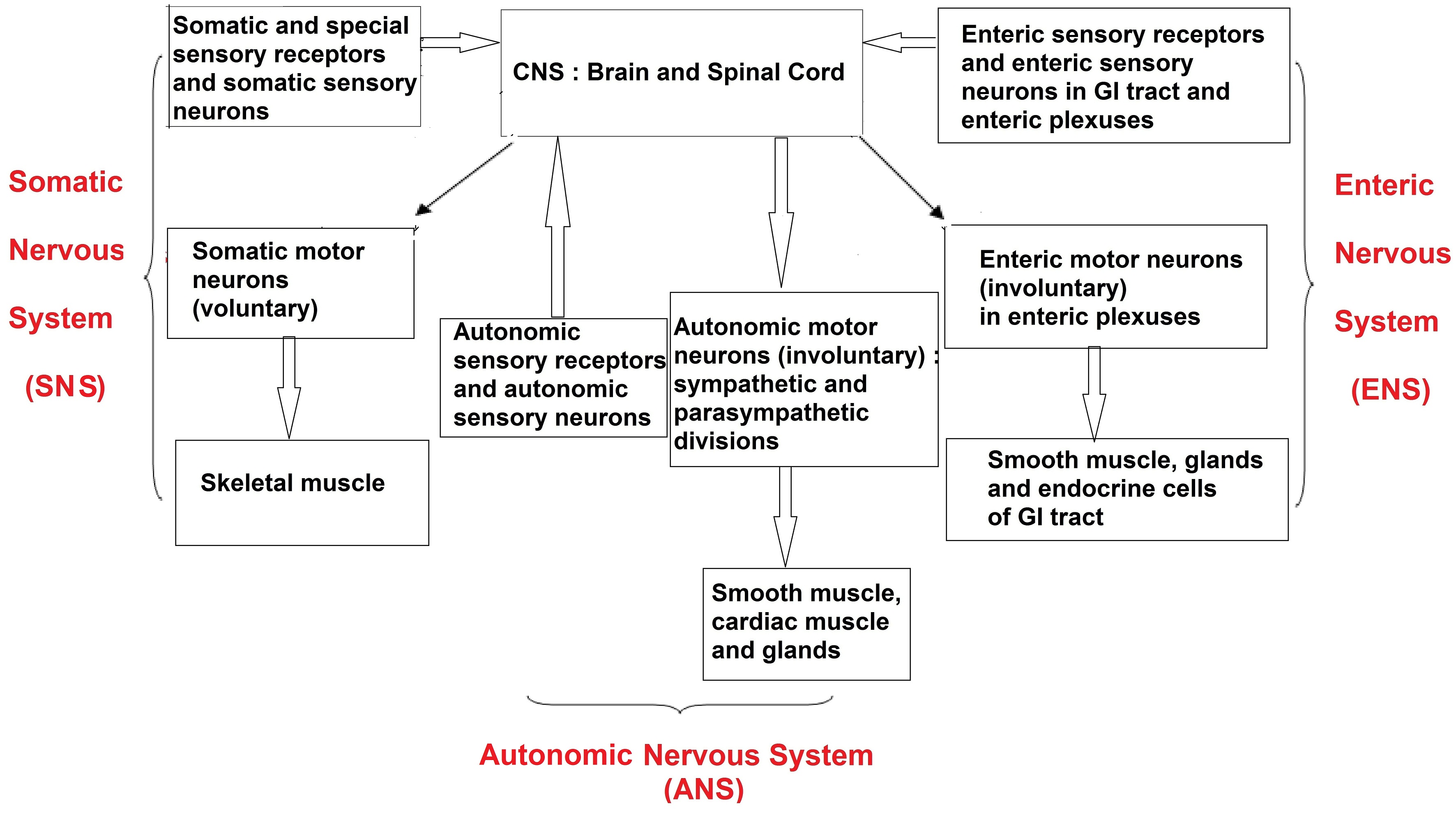
A. Somatic Nervous System (SNS) consists of –
- Sensory neurons that convey information from somatic receptors of head, trunk, limbs as well as from special senses of vision, hearing etc to the CNS.
- Motor neurons that conducts impulses from CNS to skeletal muscles only. This motor response is consciously controlled therefore (SNS) is under voluntary control.
B. Autonomic Nervous System (ANS) consists of –
- Sensory neurons, that convey information from autonomic sensory receptors which are located in visceral organs like stomach and lungs to CNS
- Motor neurons, that conduct nerve impulses from CNS to smooth muscles, glands etc. Because its motor response is not normally under conscious control it is involuntary in nature.
The motor nerves of ANS have 2 divisions.
- Sympathetic division (Fig. 6)
- Parasympathetic division (Fig. 7)
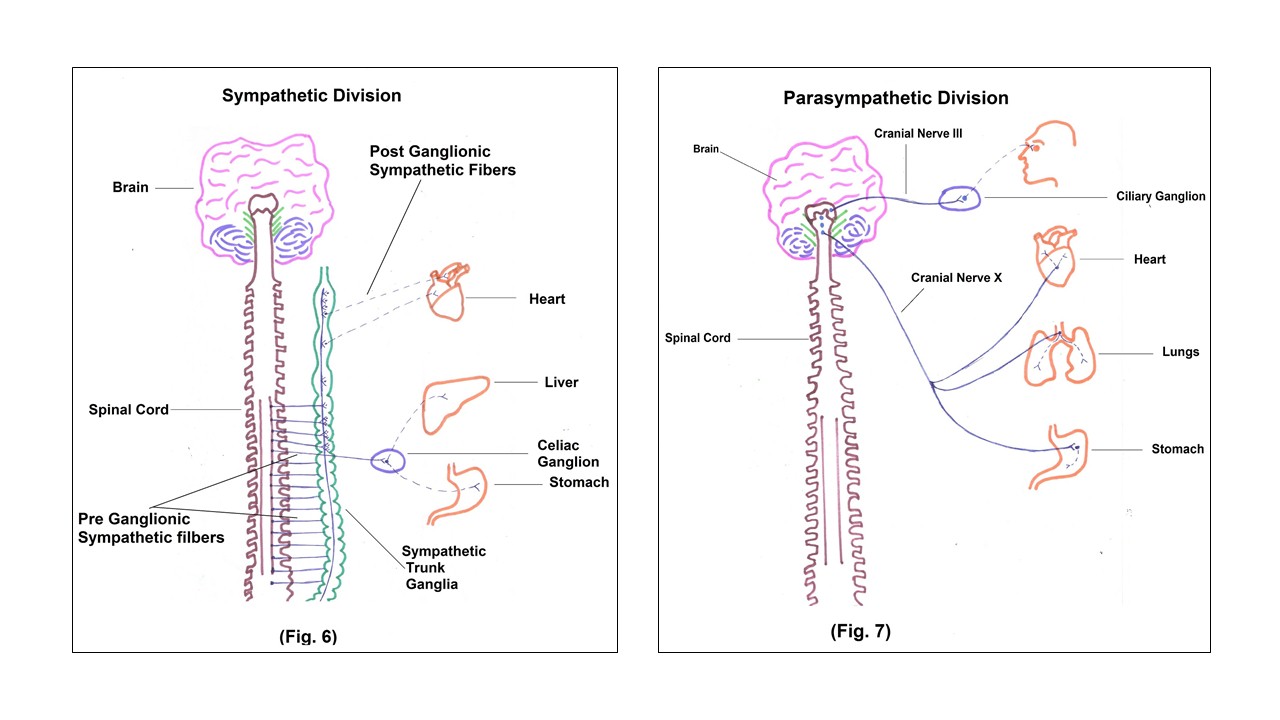
Most organs have dual innervations
One division – Excitatory
Another division – Inhibitory
Sympathetic division is active during fight or flight, means during emergency situation. Parasympathetic division is necessary for maintaining daily activities and conserving energy. Both the divisions are necessary for maintenance of Homoeostasis. We will see now protective covering of brain and spinal cord that is called as meninges.
Meninges consist of – (Fig.8)
- Outer dura matter
- Middle arachnoid matter
- Inner pia matter
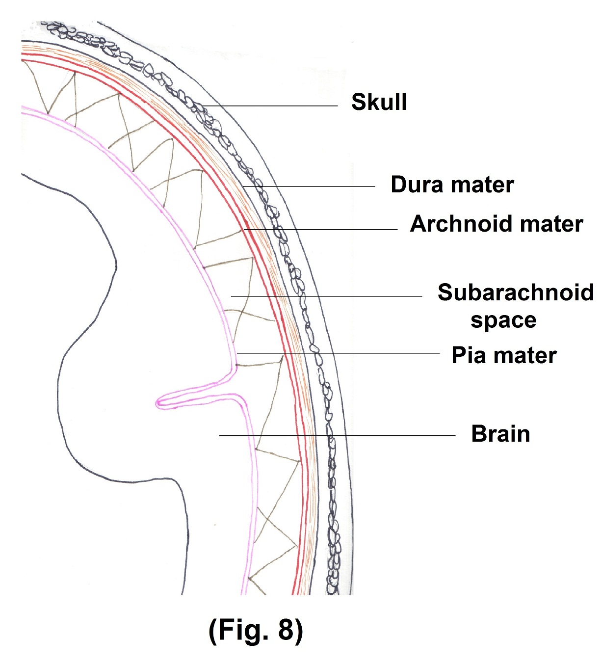
Cerebrospinal fluid, which is present in ventricles and sub arachnoid space acts as a shock absorber. It protects the brain and spinal cord from chemical and physical injuries. Rate of formation and reabsorption of CSF are same. Therefore pressure of CSF is normally constant.
Blood flow of brain —
Blood reaches brain via circle of Willis and dural venous sinus drain it into internal jugular vein to return blood from head to heart. For synthesis of ATP, neuron requires O2 therefore even brief slowing of blood flow to brain may cause disorientation or even lack of consciousness. Though brain has 2% of total body weight, it consumes about 20% of O2 and glucose used by the body. As glucose is not stored in brain, Hypoglycemia leads to confusion, dizziness, loss of consciousness and sometimes convulsions.
Blood brain barrier –
As the name suggest, this partition does not allow all substances to pass from blood into the brain. Glucose passes through blood – brain barrier easily by active transport whereas proteins and most antibiotics do not pass at all from blood into brain tissue. Trauma, certain toxins and inflammation can cause breakdown of BBB.
Let us have a few examples of working of the nervous system.
If someone touches your hand, how the message reaches the brain and how action occur in response to that? (Fig.9 – Description Nos. as below)
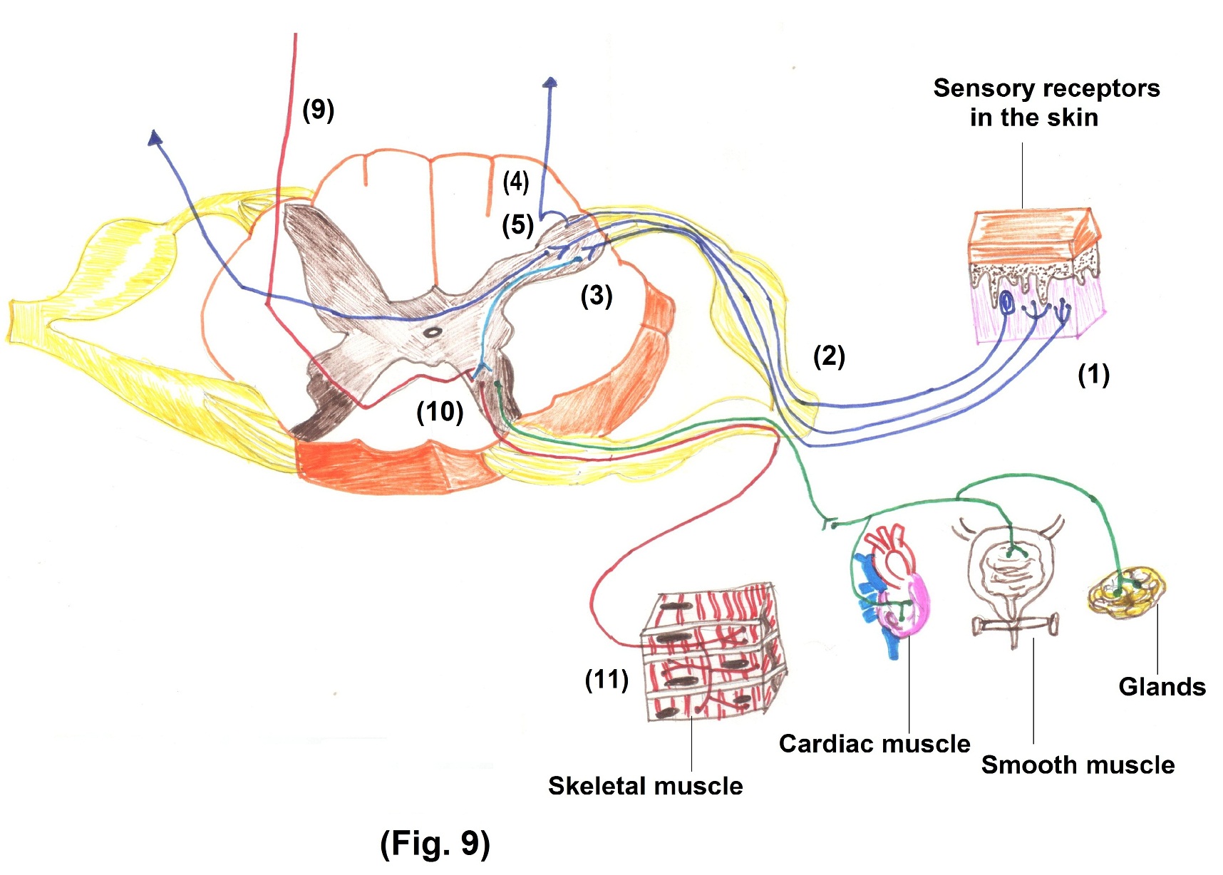
- Touch receptors on your hand detect the stimuli i.e. pressure from the touch.
- Sensory neurons convey this nerve impulse to spinal nerve and then into posterior root of spinal cord. From posterior root axons of sensory neuron may proceed along 3 possible pathways.
- Axons of sensory neuron synapse with interneuron in posterior horn and synapse with somatic motor neuron and spinal reflex occur immediately.
- Axons of sensory neurons may extend into white matter of spinal cord and ascend to the brain as part of sensory tract.
- Axons of sensory neurons cross over at the level of spinal cord and enter into posterior gray horn and then enter into white matter and ascend to the brain as part of sensory tract.
- Then this sensory ascending tract reaches to the thalamus and synapse with another neuron.
- The axon of this neuron reaches sensory area of cerebral cortex, where sensation of touch occurs.
- Then via interneuron impulse reaches to motor area of cerebral cortex.
- Some of the motor neurons cross at brain stem level. Most of the fibres are cranial nerves. Rest of the neuron cross at spinal cord level and reach the anterior root of spinal cord.
- From anterior root it enters into spinal nerve and extends to the skeletal muscles of body.
- Then contraction of skeletal muscle occur leading to appropriate movement of your hand.
Motor pathways contain
- Upper motor neurons and
- Lower motor neurons
Upper motor neurons originate in cerebral cortex and in several brain stem nuclei and their axons synapse with motor nuclei in the brain stem for cranial nerves and in spinal cord for spinal nerves.
Lower motor neurons have cell bodies in spinal cord and their axons carries impulses through spinal nerves into peripheral nerves and ending at neuromuscular junctions in muscles.
When upper motor neurons are damaged above the crossover of its tracts in the medulla, motor impairment develops on the opposite side. If damage occurs below the crossover, effects are noticed on the same side of the body. The affected limb becomes weak or paralyzed and skilled movements are performed poorly as compared to gross movements. Muscle tone is increased and deep tendon reflexes are exaggerated.
Damage to lower motor neurons causes weakness and paralysis on the same side however, muscle tone and reflexes are decreased or absent.
Let us have an example to understand the control and coordination function of the nervous system. (Fig. 10)
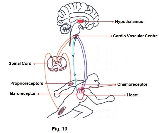
- You are going to start a running race
- Through your special senses like vision and hearing, nerve impulse reaches to brain stem and from there some axons reach to visual areas and auditory areas respectively in cerebral cortex. Thus, you become aware of the environment around you.
- Equilibrium pathway from cerebellum helps you to adjust position of your body.
- As it is a competition, concomitantly, limbic system is activated because of anticipation. Signals from limbic system are sent to cerebrum. This in turn results into nerve impulses from cerebrum to CVC (Cardiovascular Center) to increase heart rate to meet extra blood supply to body.
- When actual running starts, proprioceptors that monitor movements of limbs send nerve impulse to cardiovascular centre to increase the heart rate and also nerve impulses to equilibrium centre in medulla, pons and cerebellum to adjust head movements according to body movements with help of motor pathways.
- As pressure of blood in blood vessels alters due to the foregoing changes, baroreceptors in arch of aorta and carotid artery stimulate cardiovascular centre and increases heart rate.
- The energy requirement increases during this process. It leads to metabolic alterations, in turn stimulating chemoreceptors. Further activation of CVC takes place. Metabolic alterations involve stimulation of pituitary gland and adrenal cortex by hypothalamus. This leads to secretion of hormones like steroid hormones.
- Requirement of O2 is also more, therefore respiratory centre in medulla also gets activated and it increases respiratory rate to fulfill the requirement of O2.
As you will observe, along with Nervous system, other systems participate in the process. This is an example of integrative functioning of nervous system.