Vision and Eye
Vision is the act of seeing. The eyes are responsible for the detection of visible light. The color of visible light depends on its wavelength. (Fig. 1)
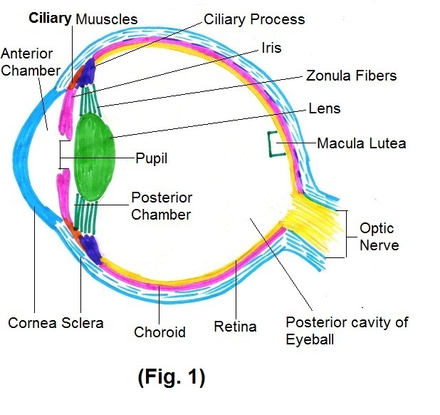
Reflected light rays from any object first fall on cornea. Cornea is a superficial layer of eyeball which is a transparent coat covering colored iris. The first refraction occurs at cornea for focusing rays on retina. Refraction means bending of light rays when they travel from one transparent medium into another. After cornea, light rays travel through anterior chamber which lies between cornea and iris. Then it travels through posterior chamber. In both, anterior and posterior chambers, slight refraction of rays occurs, and both contain aqueous humor, a transparent watery fluid that nourishes the lens and cornea. From posterior chamber light rays travel up to lens through pupils. Autonomic reflexes regulate pupil diameter in response to light. When there is bright light, it stimulates parasympathetic fibers of oculomotor nerve which in turn stimulate the circular muscles of iris. It results into contraction of circular muscles of iris, causing decrease in size of pupil. (Fig. 2)
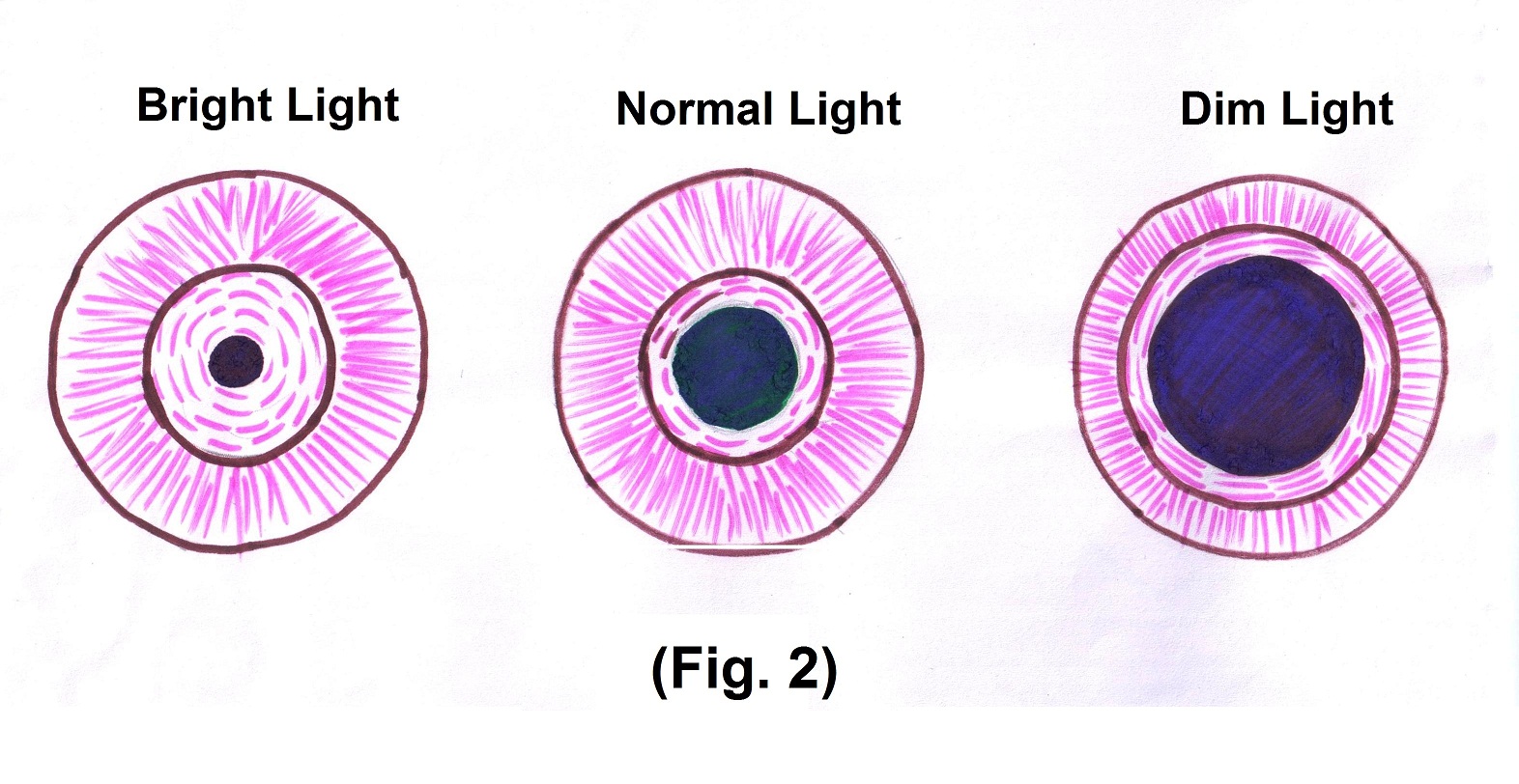
In dim light sympathetic nerves stimulate radial muscles of the iris to contract, causing an increase in the size pupil.
Then light rays reach at lens. Lens is located behind pupil and iris, and within the cavity of eyeball. The lens helps in focusing images on the retina to facilitate clear vision. It changes the shape with help of ciliary muscles, adapting it for near or far vision. In old age, lens loses elasticity causing refraction error called presbyopia. After the lens light rays enter into larger posterior cavity of eyeball, which lies between the lens and retina. It contains vitreous body, a transparent jelly like substance that holds retina giving it an even surface for the reception of clear images. Slight refraction of light rays occurs in posterior cavity. The vitreous body also contains phagocytic cells that remove debris, keeping this part of the eye clear for unobstructed vision. The pressure in the eye, intraocular pressure, is produced mainly by the aqueous humor. Increase in the pressure of aqueous humor causes glaucoma.
Ultimately light rays reach last layer of eyeball called retina. (Fig. 3)
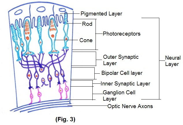
Retina consists of pigmented layer and neural layer. Pigmented layer helps to absorb light rays going in the wrong direction. The neural layer processes visual data before sending nerve impulses into axons that form the optic nerve. Neural layer has three layers
- Photoreceptor layer
- Bipolar cell layer
- Ganglion cell layer.
There are two types of photoreceptors, Rods and Cones. Rods allow us to see in dim light. Decrease in Rods causes night blindness. It occurs mostly due to vitamin A deficiency. Bright light stimulates cones which produce color vision and loss of the cones produces color blindness. Three types of Cones are present in retina, Blue cone, Green cones and Red cones. Deficiency of any one type of cone causes color blindness. Photoreceptors are specialized cells that begin the process by which light rays ultimately converted to nerve impulses.
Light rays pass through bipolar cell layer and ganglion cell layer before it reaches photoreceptor layer. In these layers modification of signals occurs. (Fig. 4)
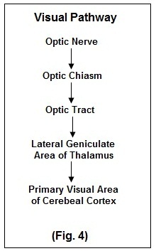
Nerve impulse from axons of optic nerve passes through optic chiasm a crossing point of optic nerves. After passing through the optic chiasm, the axons now part of optic tract, enter the brain and most of them terminate in the lateral geniculate nucleus of the thalamus. Ultimately it reaches primary visual areas in the cerebral cortex and visual perception occurs.